
Patient guide
Imaging and scans for the upper limb
Shoulder, elbow, hand and wrist problems can look alike from the outside. Mr Jepson uses the scan best suited to the question being asked, so you have the test that will genuinely help, and no more than you need.
This guide explains what each type of imaging shows, what it is like to have, and how safe it is.
Before any scan
The examination comes first
Every assessment starts with a conversation about your symptoms and a careful examination of your shoulder, elbow, hand or wrist. In many cases this points clearly towards the likely diagnosis before any scan is arranged.
Imaging is then used to answer specific questions: to confirm the diagnosis, to rule out other causes of pain, and to help plan treatment or surgery when it is needed.
Confirm
What the examination suggests
Rule out
Other causes of your symptoms
Plan
The right treatment or operation
Not everyone needs a scan, and many patients need only one. Mr Jepson will explain why a particular scan is being recommended and what the result may change.
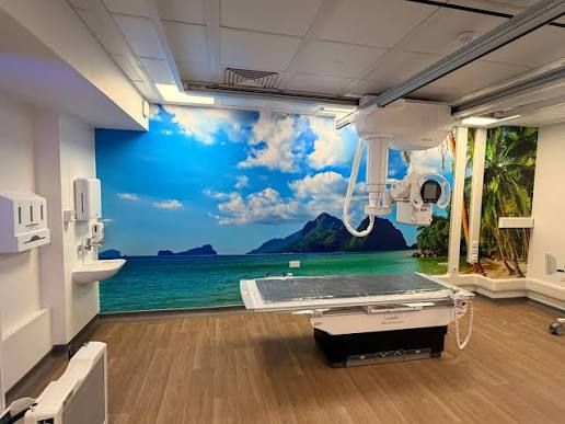
Scan 1
X-ray
An X-ray is usually the first and simplest picture of the bones and joints. It is quick, widely available and often all that is needed.
What it shows
- Bones and their alignment
- Joint space and arthritis
- Calcium deposits in tendons
- Fractures
- Existing implants and metalwork
Commonly used for
Thumb base arthritis, calcific tendonitis of the shoulder, fractures, and checking the joint after a shoulder dislocation.
Safety
X-rays are quick and painless. They use a small dose of ionising radiation, so please tell the staff if there is any chance you might be pregnant.
Scan 2
Diagnostic ultrasound
Ultrasound uses sound waves to build a real-time picture of the tendons, bursae, ligaments and any fluid around a joint. Because the image is live, structures can be watched as you move your arm, which is known as a dynamic assessment, and the painful side can be compared with the other side.
In the shoulder, it shows calcium deposits in the rotator cuff tendons, as in calcific tendonitis of the shoulder. For a lump, ultrasound confirms whether it is a fluid-filled cyst, as with ganglion cysts of the wrist and hand, and shows how it sits against nearby nerves and blood vessels.
Commonly used for
- Rotator cuff tears
- Calcific deposits in the shoulder
- Bursitis
- Biceps tendon problems
- Tendon changes in tennis elbow
- Ganglia (fluid-filled lumps)
Safety
No radiation, painless, and safe in pregnancy.
Limitations
Sound waves cannot see inside bone or deep joint structures well, so an MRI may sometimes be added.
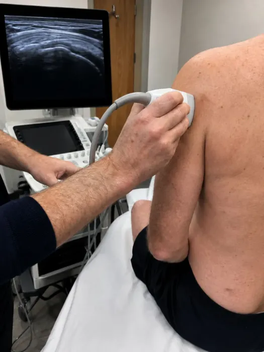
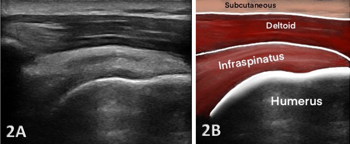
Imaging-guided treatment
Ultrasound-guided injections
Ultrasound can also guide treatment. Watching the needle on screen means it can be placed precisely into the bursa, the joint, a tendon sheath or a calcium deposit, rather than relying on landmarks felt through the skin.
Subacromial steroid injection
Into the bursa above the rotator cuff, to settle inflammation and pain.
Hydrodistension
Gently stretching the tight joint capsule in frozen shoulder.
Barbotage
Breaking up and washing out calcium in calcific tendonitis.
Carpal tunnel injection
Alongside the median nerve in carpal tunnel syndrome.
Thumb base injection
Into the small joint at the base of the thumb in thumb base arthritis.
What happens
Local anaesthetic numbs the skin first. It is a short outpatient procedure and you go home the same day.
Possible side effects
- A temporary flare of pain after the injection
- A small risk of infection
- Thinning or lightening of the skin at the injection site
- A temporary rise in blood sugar for people with diabetes
- Temporary facial flushing
When to contact the practice
If the injection site becomes red, hot or swollen, or you develop a fever, please contact the practice promptly so this can be checked.
Call 01604 633730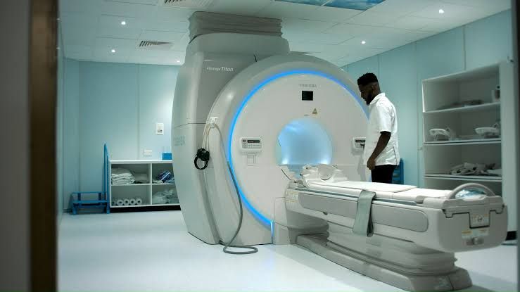
Scan 4
MRI
MRI uses a strong magnet and radio waves, with no ionising radiation, to produce detailed pictures of the soft tissues inside and around a joint.
Best for
Tendons, cartilage, ligaments and nerves, bone bruising, and hidden fractures that may not show on X-ray, such as a fracture of the scaphoid bone in the wrist.
Commonly used for
Rotator cuff tears and planning cuff repair, cartilage problems, and wrist ligament injuries.
What it is like
It takes longer than an X-ray. You lie still while the scanner makes loud knocking sounds, and ear protection is provided.
Safety screening
You will be asked about pacemakers, some implants and any metal fragments. Please mention claustrophobia in advance.
Open and upright MRI
A standard MRI scanner is a narrow tunnel. Some people find this difficult, for example because of claustrophobia, a larger build, or pain that makes it hard to lie still in a set position for the length of the scan.
Open MRI
The scanner has open sides and more space around your body, which many people find easier and less enclosed.
Upright MRI
Some scans can be taken while you sit or stand, so a joint can be imaged in the position, or with the movement, that brings on your symptoms.
Open and upright scanners can produce different image detail from a standard scanner, so they are not the right choice for every question. Mr Jepson will advise whether one suits the scan you need.
Please check cover with your insurer before booking an open or upright scan.
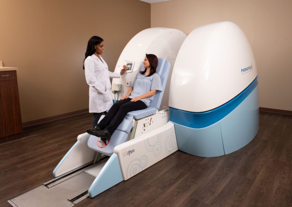
Nearest scanners
Avicenna Clinic, Peterborough
An open MRI scanner with more space around the body.
Visit the Avicenna Clinic MRI page (opens in a new tab)Open MRI, Birmingham and London
An upright scanner, where some scans can be taken sitting or standing.
Visit the Upright MRI website (opens in a new tab)These are independent centres, and the practice team can explain how to arrange a scan there and send the images to Mr Jepson ahead of your appointment.
Scan 5
MR arthrogram
A small amount of contrast dye (gadolinium) is injected into the joint under X-ray or ultrasound guidance, and an MRI scan follows soon after. The fluid gently distends the joint, so small tears that might be missed on a standard MRI show up clearly.
Commonly used for
- Labral and SLAP tears
- Shoulder instability
- Small partial tendon tears
- Wrist ligament and TFCC tears
What it is like
There is brief discomfort from the injection, then the MRI itself as described above.
Safety
A small risk of infection and a rare allergy to the contrast. Tell staff about kidney problems or allergies.
Scan 6
CT scan
A CT scanner combines many X-ray images into detailed cross-sections of bone, which can also be turned into 3D reconstructions. It gives the clearest view of bone shape and structure.
Safety
CT uses a higher radiation dose than a plain X-ray, so it is only used when the extra detail will change your treatment. Please tell staff if you might be pregnant.
Commonly used for
-
Complex fractures
Of the shoulder, elbow and wrist, to plan how the bone fragments are best repaired.
-
Scaphoid fractures and non-union
Checking whether a wrist fracture has healed.
-
Glenoid bone loss and Latarjet planning
Measuring bone lost from the shoulder socket after repeated dislocations, and planning a Latarjet procedure. Read more in the shoulder instability guide.
-
Before shoulder replacement
Assessing the bone so the implant can be positioned accurately.
Side by side
Choosing the right scan
A simple comparison. Mr Jepson will recommend the scan that best answers the question about your symptoms.
If you find a standard MRI scanner difficult, open or upright MRI may be an option. Read about open and upright MRI.
-
X-ray
- Best for
- Bones, arthritis, calcium, fractures
- Radiation
- Yes, small dose
- Common uses
- Thumb base arthritis, calcific tendonitis, dislocation
-
Ultrasound
- Best for
- Tendons, bursae, movement in real time
- Radiation
- No
- Common uses
- Rotator cuff, bursitis, tennis elbow, ganglia
-
Ultrasound-guided injection
- Best for
- Accurate placement of treatment
- Radiation
- No
- Common uses
- Subacromial injection, hydrodistension, barbotage
-
MRI
- Best for
- Tendons, cartilage, ligaments, nerves, hidden fractures
- Radiation
- No
- Common uses
- Rotator cuff tears, wrist ligament injuries
-
MR arthrogram
- Best for
- Small tears inside the joint
- Radiation
- No, unless the injection is X-ray guided
- Common uses
- Labral and SLAP tears, TFCC tears
-
CT
- Best for
- Detailed bone and 3D reconstructions
- Radiation
- Yes, higher dose
- Common uses
- Complex fractures, glenoid bone loss, Latarjet planning
| Scan | Best for | Radiation | Common upper limb uses |
|---|---|---|---|
| X-ray | Bones, arthritis, calcium, fractures | Yes, small | Thumb base arthritis, calcific tendonitis, dislocation |
| Ultrasound | Tendons, bursae, movement in real time | No | Rotator cuff, bursitis, tennis elbow, ganglia |
| Guided injection | Accurate placement of treatment | No | Subacromial injection, hydrodistension, barbotage |
| MRI | Tendons, cartilage, ligaments, nerves, hidden fractures | No | Rotator cuff tears, wrist ligament injuries |
| MR arthrogram | Small tears inside the joint | No, unless X-ray guided | Labral and SLAP tears, TFCC tears |
| CT | Detailed bone and 3D reconstructions | Yes, higher | Complex fractures, glenoid bone loss, Latarjet planning |
Preparing
Before your scan
Please let the imaging team know about any of the following before your appointment. It helps them choose the safest approach for you.
- You are, or might be, pregnant
- You have a pacemaker or another implanted device
- You have metal fragments or have had previous surgery
- You have kidney problems
- You have had an allergic reaction to contrast dye
- You have diabetes and are having a steroid injection
- You feel anxious in enclosed spaces (claustrophobia)
- You take blood-thinning medicines and are having an injection
- Bring any previous scans or reports, including those done at another hospital
Questions
Frequently asked questions
Who reports my scan?
Your scan is reported by a consultant radiologist. Mr Jepson then reviews the images himself and discusses the results with you, explaining what they mean for your treatment. You can read more about the team he works with on the About page.
Will my insurer cover the scan?
Most insurers cover scans that your consultant recommends, but policies differ. Please check with your insurer before the scan and obtain an authorisation code.
Where are scans done?
Scans are arranged at Circle Three Shires Hospital in Northampton and Woodland Hospital in Kettering. The practice will confirm where your scan will take place. Addresses and directions are on the clinic locations page.
I am claustrophobic. Can I still have an MRI?
Usually, yes. Please mention it when your scan is booked. The radiographers can often help, for example by explaining each step beforehand and staying in contact with you throughout the scan. If a standard scanner still feels too difficult, open or upright MRI may be an alternative that Mr Jepson can discuss with you. Read about open and upright MRI.
Can I bring scans done elsewhere?
Yes. Please bring the images and the written reports, or ask the hospital that performed them to send them in advance. This can avoid repeating a scan unnecessarily.
Can I share scans I have had elsewhere?
Yes. You can upload the reports through your secure Carebit patient dashboard (opens in a new tab). Full MRI or CT image sets are often too large to upload, so the practice team can arrange a transfer between hospitals. Reports of scans Mr Jepson requests appear in your dashboard once he has reviewed them. How the patient portal works, in the patient FAQs.
Do I need a scan before my first appointment?
Not usually. Mr Jepson prefers to examine you first and then arrange the scan that best answers the question, which can sometimes be done on the same day.
Talk to the practice about your shoulder, elbow, hand or wrist
Call to arrange a consultation, book online for Circle Three Shires Hospital in Northampton, or send a short email and the practice team will come back to you.
Read independent patient reviews on Doctify (opens in a new tab), iWantGreatCare (opens in a new tab) or Google reviews (opens in a new tab)